A New Era of Dental Excellence
By 2025, digital tools have become routine in most U.S. dental offices. Intra‑oral scanners now replace conventional putty impressions, delivering accurate 3‑D models in seconds and eliminating gag‑inducing materials. CAD/CAM systems such as CEREC enable same‑day design, milling, and placement of crowns, bridges, veneers and onlays, cutting turnaround time from weeks to a single visit. Integrated cone‑beam computed tomography (CBCT) and digital radiography provide high‑resolution, low‑radiation 3‑D imaging for implant planning, orthodontic assessment, and early disease detection.
These technologies dramatically improve patient comfort—digital impressions are painless, digital X‑rays reduce radiation exposure by up to 90%, and laser‑assisted procedures minimize bleeding and post‑operative pain. Clinical efficiency rises as digital workflows streamline diagnostics, treatment planning, and appliance fabrication, reducing chair time by 15‑25 % and appointments the number of visits. AI‑driven image analysis and treatment‑planning software further enhance diagnostic accuracy and personalize care.
The 2025 technology landscape is characterized by seamless integration of intra‑oral scanning, CAD/CAM milling, 3‑D printing, AI analytics, and teledentistry within unified practice‑management platforms, delivering faster, more precise, and patient‑centered dental care.
Understanding Digital Dentistry
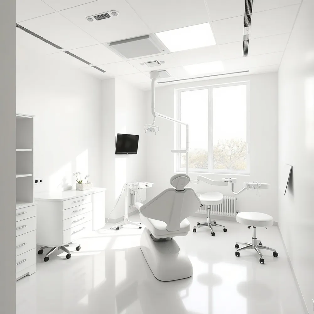
![]() Digital dentistry is the integration of computer‑based hardware and software into every stage of dental care, replacing many manual and analog processes. It utilizes tools such as intra‑oral scanners, cone‑beam computed tomography (CBCT), CAD/CAM design, 3‑D printing, digital radiography, and artificial‑intelligence‑driven treatment planning to capture, model, and fabricate restorations, orthodontic appliances, implants, and more. By creating accurate, virtual 3‑D representations of a patient’s dentition, clinicians can diagnose problems, simulate outcomes, and communicate plans with patients before any physical work begins. This digital workflow speeds up production, reduces material waste, improves precision and aesthetic quality of restorations, and enhances overall practice efficiency, leading to greater patient confidence and a smoother, more personalized experience.
Digital dentistry is the integration of computer‑based hardware and software into every stage of dental care, replacing many manual and analog processes. It utilizes tools such as intra‑oral scanners, cone‑beam computed tomography (CBCT), CAD/CAM design, 3‑D printing, digital radiography, and artificial‑intelligence‑driven treatment planning to capture, model, and fabricate restorations, orthodontic appliances, implants, and more. By creating accurate, virtual 3‑D representations of a patient’s dentition, clinicians can diagnose problems, simulate outcomes, and communicate plans with patients before any physical work begins. This digital workflow speeds up production, reduces material waste, improves precision and aesthetic quality of restorations, and enhances overall practice efficiency, leading to greater patient confidence and a smoother, more personalized experience.
What does digital dentistry do? It captures high‑resolution 3‑D images of teeth and jaws, allowing precise diagnosis and customized treatment planning. CAD/CAM and 3‑D printing then fabricate crowns, bridges, and other restorations often in a single appointment, cutting laboratory time and reducing patient visits. Digital impressions eliminate messy silicone trays, improving comfort and reducing chair time. Integrated electronic health records and practice‑management software streamline scheduling, billing, and data sharing, further enhancing efficiency.
What are the innovations in digital dentistry? Recent advances include ultra‑fast intra‑oral scanners that produce color‑accurate 3‑D models in seconds, AI algorithms that automatically detect caries, periodontal bone loss, and early oral cancer on radiographs, and generative design platforms that suggest optimal restorative shapes. 3‑D printing now offers biocompatible ceramic and resin materials for same‑day prosthetics, while laser‑assisted procedures provide minimally invasive tissue removal with less pain and faster healing. Teledentistry platforms expand access by enabling remote consultations, follow‑up care, and patient education, making modern dental care more convenient and patient‑centered.
Digital Workflow in Practice
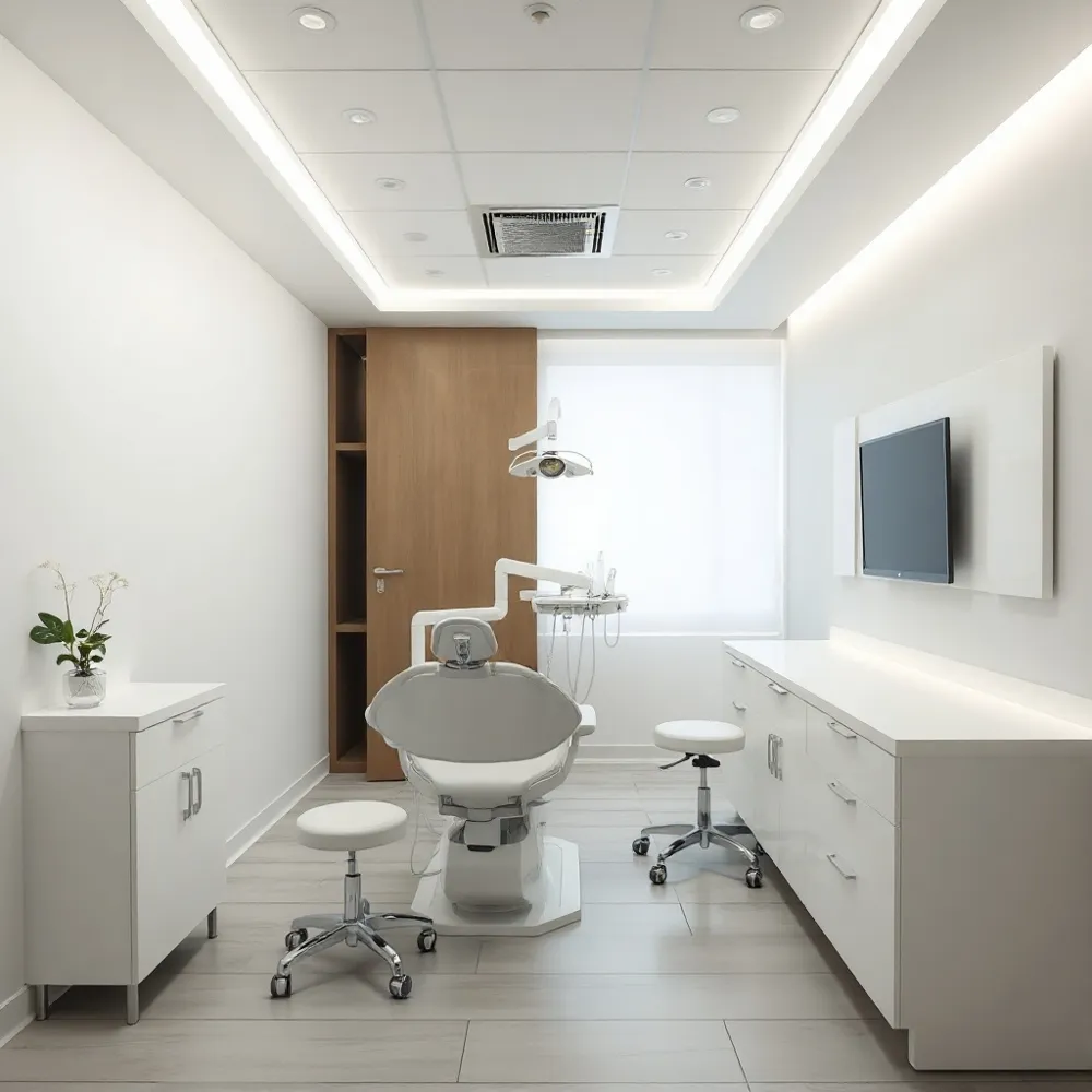
![]() Integration of imaging, CAD design, and CAM manufacturing streamlines practice operations. Digital X‑rays and CBCT deliver high‑resolution, low‑radiation imaging that feed directly into the design workflow, eliminating manual data entry. Practice‑management software links patient records, appointment scheduling, and communication tools, allowing clinicians to send treatment simulations to patients for informed consent and to receive real‑time feedback. This seamless data flow reduces chair time, cuts the number of visits, and improves resource utilization.
Integration of imaging, CAD design, and CAM manufacturing streamlines practice operations. Digital X‑rays and CBCT deliver high‑resolution, low‑radiation imaging that feed directly into the design workflow, eliminating manual data entry. Practice‑management software links patient records, appointment scheduling, and communication tools, allowing clinicians to send treatment simulations to patients for informed consent and to receive real‑time feedback. This seamless data flow reduces chair time, cuts the number of visits, and improves resource utilization.
What is a digital workflow in dentistry? A digital workflow is a paper‑free, end‑to‑end process that captures patient anatomy with digital scans, plans treatment virtually, designs restorations via CAD, manufactures them with CAM, and integrates all information with the practice’s electronic health record (EHR) and communication systems, enhancing accuracy, efficiency, and patient experience.
What are the benefits of digital dentistry? It offers superior precision through 3‑D imaging and intra‑oral scanners, enables same‑day design and fabrication of restorations, reduces patient discomfort by eliminating traditional impressions, supports early disease detection, and lowers material waste and laboratory costs, resulting in faster, more personalized, and cost‑effective care.
Digital Prosthodontics & Implant Guidance
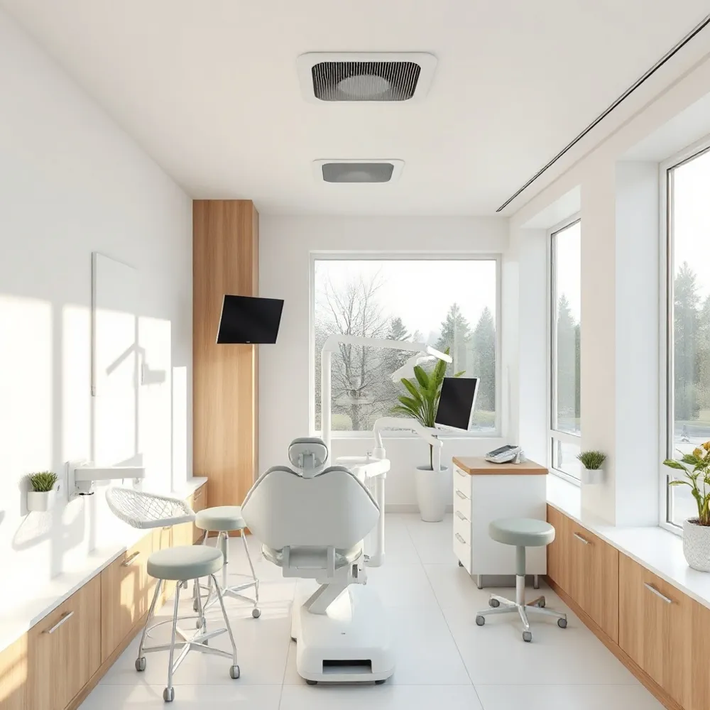
![]() Digital dentistry has reshaped prosthodontics by replacing conventional impressions with intra‑oral scanners that capture highly accurate 3‑D models of the dentition and surrounding soft tissues. These digital files are imported into CAD software where clinicians design crowns, bridges, dentures, and implant‑supported prostheses with precise margins, occlusal contacts, and aesthetic contours. The completed designs are fabricated on‑site by CAM units—either high‑precision milling machines or 3‑D printers—using biocompatible ceramic or resin materials that deliver superior fit and strength. Because the workflow is fully digital, many restorations can be completed in a single appointment, eliminating temporary crowns and reducing chair‑time, which translates into higher patient satisfaction.
Digital dentistry has reshaped prosthodontics by replacing conventional impressions with intra‑oral scanners that capture highly accurate 3‑D models of the dentition and surrounding soft tissues. These digital files are imported into CAD software where clinicians design crowns, bridges, dentures, and implant‑supported prostheses with precise margins, occlusal contacts, and aesthetic contours. The completed designs are fabricated on‑site by CAM units—either high‑precision milling machines or 3‑D printers—using biocompatible ceramic or resin materials that deliver superior fit and strength. Because the workflow is fully digital, many restorations can be completed in a single appointment, eliminating temporary crowns and reducing chair‑time, which translates into higher patient satisfaction.
Guided implant surgery builds on this workflow: CBCT imaging and intra‑oral scans are merged to create a virtual treatment plan, and a 3‑D‑printed surgical guide is produced to direct the drill to the exact planned position. This integration improves placement accuracy, minimizes surgical risk, and enhances the predictability of the final prosthetic outcome.
How is digital dentistry used in prosthodontics? Digital scanning, CAD design, and CAM fabrication enable precise, single‑visit restorations with better fit, reduced discomfort, and instant data for future replacements.
Why don't dentists do fillings anymore? Amalgam fillings are being phased out due to mercury concerns and aesthetic limitations; modern composite resins bond directly to tooth structure, preserving healthy tissue and providing a tooth‑colored, durable restoration.
Digital Dentures: Care, Eating & Longevity
![]() CAD/CAM‑fabricated digital dentures are milled from high‑strength, biocompatible resin, providing a precise fit that mimics natural tooth anatomy and distributes occlusal forces evenly. Their accuracy reduces sore spots and improves chewing efficiency compared with traditional acrylic dentures.
CAD/CAM‑fabricated digital dentures are milled from high‑strength, biocompatible resin, providing a precise fit that mimics natural tooth anatomy and distributes occlusal forces evenly. Their accuracy reduces sore spots and improves chewing efficiency compared with traditional acrylic dentures.
Dietary recommendations after placement – In the first 24–48 hours patients should stick to soft, easy‑to‑chew foods such as soups, smoothies, oatmeal, scrambled eggs, and well‑cooked vegetables. As comfort increases, firmer items can be introduced in small, bite‑size pieces and cooked until tender. Hard, sticky, or crunchy foods (hard nuts, popcorn kernels, hard candy, chewing gum, raw carrots, tough meats) should be avoided because they can crack or dislodge the prosthesis. Very hot foods should be allowed to cool slightly to prevent warping of the resin.
Expected lifespan and maintenance – Digital dentures typically last 5–10 years when properly cared for, comparable to conventional dentures. Their durability is supported by twice‑daily brushing with a soft‑bristled brush, non‑abrasive toothpaste, and routine dental check‑ups. Implant‑supported digital dentures can extend the prosthetic’s functional period, with the implant portion lasting a lifetime while the denture superstructure may need replacement every 10–15 years.
Can you eat with digital dentures? Yes—you can eat with digital (CAD/CAM) dentures, but start with soft foods and gradually re‑introduce firmer items while avoiding extremely hard, sticky, or very hot foods.
How long do digital dentures last? With diligent cleaning and regular professional visits, they usually remain functional for 5–10 years; implant‑supported versions may last even longer, though the denture itself may still require periodic replacement.
Patient‑Centred Hygiene & Aesthetic Rules
![]() For a harmonious smile, the 50‑40‑30 rule guides contact‑area proportions among the upper front teeth. The central‑central contact should cover roughly 50 % of the tooth length, the central‑lateral contact about 40 %, and the lateral‑canine contact about 30 %. This graduated reduction creates a youthful, balanced appearance and minimizes black‑triangle spaces. Apply the rule when planning veneers, crowns, or full smile makeovers, adjusting percentages to each patient’s anatomy.
For a harmonious smile, the 50‑40‑30 rule guides contact‑area proportions among the upper front teeth. The central‑central contact should cover roughly 50 % of the tooth length, the central‑lateral contact about 40 %, and the lateral‑canine contact about 30 %. This graduated reduction creates a youthful, balanced appearance and minimizes black‑triangle spaces. Apply the rule when planning veneers, crowns, or full smile makeovers, adjusting percentages to each patient’s anatomy.
Practical tip: combine digital smile‑design software with these rules to visualize outcomes, enhance patient education, and ensure both oral health and aesthetic goals are met.
Future Outlook: AI, 3‑D Printing & Tele‑Dentistry
![]() Artificial intelligence will soon move beyond image analysis to predictive treatment planning, using machine‑learning models that combine CBCT scans, intra‑oral scans and patient histories to forecast caries risk, optimal implant positioning and orthodontic outcomes. 3‑D printing is entering a new era as resin and ceramic powders become cheaper and biocompatible, enabling on‑site fabrication of high‑precision crowns, surgical guides and even bio‑active scaffolds at a fraction of today’s cost. Tele‑dentistry platforms, bolstered by secure cloud‑based EHRs, will extend these diagnostics to rural and underserved communities, allowing remote imaging review, AI‑assisted triage and virtual treatment simulations. Emerging adjuncts—laser‑assisted soft‑tissue work, AR/VR visualizations for patient education, and robotic‑assisted implant placement—will further streamline workflows, improve accuracy and enhance the patient experience.
Artificial intelligence will soon move beyond image analysis to predictive treatment planning, using machine‑learning models that combine CBCT scans, intra‑oral scans and patient histories to forecast caries risk, optimal implant positioning and orthodontic outcomes. 3‑D printing is entering a new era as resin and ceramic powders become cheaper and biocompatible, enabling on‑site fabrication of high‑precision crowns, surgical guides and even bio‑active scaffolds at a fraction of today’s cost. Tele‑dentistry platforms, bolstered by secure cloud‑based EHRs, will extend these diagnostics to rural and underserved communities, allowing remote imaging review, AI‑assisted triage and virtual treatment simulations. Emerging adjuncts—laser‑assisted soft‑tissue work, AR/VR visualizations for patient education, and robotic‑assisted implant placement—will further streamline workflows, improve accuracy and enhance the patient experience.
Embracing the Digital Smile
Digital dentistry has revolutionized oral care by delivering precise 3‑D imaging, intra‑oral scanning, CAD/CAM same‑day restorations, and AI‑driven diagnostics, which together shorten appointments, lower radiation, and boost patient confidence. In Midland, Texas, Dr. Ashley E. Burns, DDS blends intra‑oral scanners, CBCT, CEREC milling, and 3‑D‑printed guides into a seamless workflow that personalizes every treatment plan and lets patients view their future smile in real time. Experience this cutting‑edge approach—schedule your tech‑enabled consultation today. Our friendly team ensures comfort, education, and coordination throughout each visit.