A New Era in Diagnostic Imaging
Digital X-rays represent a transformative advancement in medical and dental imaging technology, offering safer, faster, and clearer diagnostic tools compared to traditional X-ray methods. This article explores the foundational technology behind digital X-rays, their advantages in patient safety and image clarity, safety standards, and the diverse applications across healthcare and industry. We also address common misconceptions and look ahead to future innovations shaping the field.
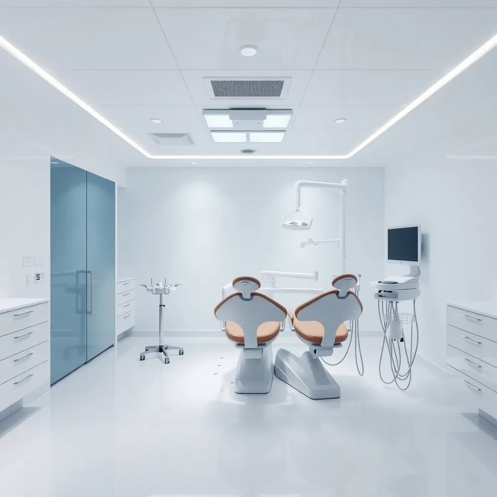
The Technology Behind Digital X-Rays: From Sensors to Software
![]()
What are the basic principles and technology behind digital X-rays, and how do they differ from traditional X-rays?
Digital X-ray technology is based on electronic sensors called detectors that directly convert X-ray photons into digital signals. This process enables immediate image acquisition, manipulation, and sharing, unlike traditional X-rays which rely on photographic film that requires chemical development, often taking minutes or hours. Digital systems utilize advanced components such as flat panel detectors equipped with materials like amorphous selenium or cesium iodide, which efficiently capture X-ray energy. The electronic nature of digital X-rays allows for lower radiation doses—up to 90% less—while producing clearer, more detailed images. These images can be quickly enhanced through software adjustments like contrast and brightness, greatly improving diagnostic accuracy and patient safety. For more information on Understanding Digital X-Rays and Advantages of Digital X-rays.
Types and mechanisms of digital X-ray detectors
Digital detectors are primarily classified into indirect and direct conversion types. Indirect detectors use scintillators such as cesium iodide or gadolinium oxysulfide that convert X-ray photons into visible light, which is then detected by a CCD or CMOS sensor. This process involves an intermediate step, which can sometimes result in slight image blurring but offers good spatial resolution.
Direct detectors, on the other hand, use amorphous selenium or cadmium telluride materials that convert X-ray photons directly into electrical charges without light intermediates. This direct process allows for sharper, higher-resolution images, especially valuable in applications like mammography. Further technical details can be found in Direct vs Indirect Digital Radiography and Digital Radiography Overview.
Image processing and enhancement techniques
Once captured, digital images can be processed using sophisticated software. This includes contrast adjustment, magnification, noise reduction, and artifact correction, all of which improve image clarity and diagnostic capabilities. Automated algorithms utilize deep learning and AI to further analyze images, highlight abnormalities, and support healthcare providers in making accurate decisions. Explore AI integrations and image enhancements at Improving Clarity and Focus for Radiologists in X-ray Imaging and AI Dental Imaging vs Digital X-rays.
Workflow improvements and electronic image management
Digital X-rays streamline workflows by enabling instant image viewing, reducing the need for physical storage and chemical processing. Images are stored securely in electronic health records and can be easily shared with specialists, facilitating remote diagnosis and collaboration. Modern digital radiography also supports integration with Picture Archiving and Communication Systems (PACS), improving overall efficiency and patient care. Learn more about Digital Radiography Workflow Improvements and Digital Radiography Image Sharing and Storage.
This combination of advanced sensors, hardware, and software innovations makes digital X-ray technology a vital tool in modern medicine, providing safer, faster, and more precise diagnostic imaging compared to traditional methods. For a comprehensive perspective, see Advantages of Digital Radiography and Digital X-rays vs Traditional X-rays.
Enhanced Safety and Diagnostic Precision with Digital X-Rays
![]() Digital X-rays have revolutionized medical and dental imaging by significantly improving safety and diagnostic accuracy. One of the foremost advantages is the drastic reduction in radiation exposure. These systems emit up to 90% less radiation than traditional film X-rays, greatly decreasing the risk of radiation-linked health issues, including cancer. Such safety improvements are especially vital for vulnerable groups like children and pregnant women, who require minimal exposure (Digital X-rays safety benefits, Digital X-rays safety in Naperville).
Digital X-rays have revolutionized medical and dental imaging by significantly improving safety and diagnostic accuracy. One of the foremost advantages is the drastic reduction in radiation exposure. These systems emit up to 90% less radiation than traditional film X-rays, greatly decreasing the risk of radiation-linked health issues, including cancer. Such safety improvements are especially vital for vulnerable groups like children and pregnant women, who require minimal exposure (Digital X-rays safety benefits, Digital X-rays safety in Naperville).
In addition to safety, digital X-rays offer superior image clarity. Their high-resolution digital sensors allow for detailed visualization of structures. Physicians and dentists can adjust contrast, brightness, magnification, and even color settings on the images, which enhances the detection of cavities, fractures, bone loss, and tumors (Digital X-rays identifying cavities and bone loss, Digital X-ray technology accuracy improvements). This level of precision not only leads to more accurate diagnoses but also facilitates early intervention.
Immediate image availability is another critical benefit. Digital systems provide real-time images directly on computer screens, enabling quick review and interaction with patients. This instant feedback accelerates diagnosis and treatment planning, especially in emergency or urgent care scenarios (Advantages of Digital X-ray, Digital X-rays speed emergency treatment). Moreover, digital images are easily stored, retrieved, and shared electronically, promoting seamless communication among healthcare teams and supporting telemedicine initiatives.
Beyond clinical benefits, digital radiography contributes positively to environmental sustainability. The technology eliminates the need for chemical processing of films, reducing hazardous waste. Digital images require less physical storage space and minimize the use of toxic chemicals associated with traditional film development.
Overall, digital X-rays combine decreased radiation risk with enhanced image quality and operational efficiency. They support safer, more accurate, and faster diagnostics, ultimately improving patient outcomes and streamlining healthcare workflows (Advantages of Digital Radiography, Benefits of digital X-rays).
Comparative Advantages Over Conventional X-Ray Systems
![]() Digital X-rays have revolutionized medical and dental imaging by offering numerous benefits that surpass traditional X-ray techniques.
Digital X-rays have revolutionized medical and dental imaging by offering numerous benefits that surpass traditional X-ray techniques.
One of the most important improvements is the reduction in radiation dose. Digital systems typically expose patients to more than 75% less radiation than conventional film X-rays. For example, where traditional X-rays might deliver around 0.005 millisieverts per image, digital X-rays often stay well below this level, significantly decreasing the risk of radiation-related health issues, especially important for frequent or pediatric imaging (Digital X-rays safety benefits, Digital X-rays for children and frequent patients).
In terms of image quality, digital X-rays provide much clearer, high-resolution images than traditional X-ray films. They offer enhanced contrast and the ability to manipulate images—increasing contrast, brightness, or magnification—allowing healthcare professionals to detect subtle abnormalities such as small fractures, early caries, or soft tissue inconsistencies with greater accuracy (Clarity and detail in Digital X-Rays, Digital X-ray technology accuracy improvements.
Environmental and operational benefits are also notable. Digital systems eliminate the need for chemical developers and film storage, which not only reduces costly chemical waste but also minimizes environmental pollution. The absence of chemicals speeds up workflows and reduces setup and cleanup times, increasing overall efficiency (Environmental benefits of digital X-rays, Digital radiography evolution and benefits).
Cost considerations favor digital X-rays over time. Although initial investment in digital equipment can be higher, operational costs decrease due to lower expenses on film, chemicals, and storage. Digital images facilitate faster diagnosis and treatment planning, leading to improved workflow and patient turnover (Cost comparison of digital and conventional X-ray systems, Workflow improvements with digital X-rays).
However, some challenges remain, notably ensuring proper dose management. Overexposure can occur if practitioners rely excessively on the capabilities of digital imaging, which appears more forgiving. Continuous training and quality control are essential to maintain optimal radiation doses while benefiting from the superior image quality and operational efficiency (Dose management and training in digital radiography, Digital radiography training and quality control).
In summary, digital X-ray technology offers substantial advantages over conventional systems, including reduced radiation risk, better image clarity, environmental friendliness, cost-effectiveness over the long term, and smoother workflow, positioning it as the standard in modern diagnostic imaging.
Applications and Future Directions in Digital Radiography
![]() Digital X-rays have revolutionized imaging in various sectors, including medical, dental, veterinary, and industrial fields. Their widespread application is driven by superior image quality, fast processing times, and enhanced safety due to lower radiation exposure. In medicine, digital X-rays facilitate the rapid diagnosis of conditions such as lung infections, fractures, and soft tissue abnormalities. They often incorporate AI-driven analysis tools that assist radiologists by detecting anomalies like tumors or lesions and providing objective, consistent diagnostics (AI dental imaging). Advanced 3D imaging techniques, such as tomosynthesis, further enhance the detail and accuracy of internal views (Digital radiography overview).
Digital X-rays have revolutionized imaging in various sectors, including medical, dental, veterinary, and industrial fields. Their widespread application is driven by superior image quality, fast processing times, and enhanced safety due to lower radiation exposure. In medicine, digital X-rays facilitate the rapid diagnosis of conditions such as lung infections, fractures, and soft tissue abnormalities. They often incorporate AI-driven analysis tools that assist radiologists by detecting anomalies like tumors or lesions and providing objective, consistent diagnostics (AI dental imaging). Advanced 3D imaging techniques, such as tomosynthesis, further enhance the detail and accuracy of internal views (Digital radiography overview).
Dentistry extensively benefits from digital radiography through detailed visualization for cavity detection, implant planning, and orthodontic assessments (Digital dental X-rays benefits). In veterinary medicine, digital X-rays allow quick and precise evaluation of animal health, minimizing stress and discomfort for patients (5 advantages of digital radiography every dvm needs to know). Industrial uses include non-destructive testing (NDT) of materials, security screening, and quality assurance. These applications leverage dual-energy imaging, automatic image stitching, and digitally enhanced analysis to detect defects or foreign objects efficiently (Digital radiography overview.
Looking toward the future, digital radiography continues to evolve with several key trends. The integration of artificial intelligence promises to improve image quality, automate interpretative workflows, and optimize radiation doses (AI dental imaging. AI-assisted software can identify subtle abnormalities and support clinical decision-making, reducing diagnostic variability (Improving clarity and focus in X-ray imaging).
Technological advancements are also focused on portable, wireless, and handheld detectors, making imaging accessible at the point of care or in field conditions (Advantages of digital radiography). Such innovations enhance patient comfort and expand reach, especially in remote or resource-limited settings. Cloud-based image storage and sharing platforms enable instant access and remote consultations, fostering Telemedicine and collaborative diagnostics (Digital image sharing).
Emerging detector technologies aim for higher sensitivity, sharper resolution, and broader dynamic range, improving the clarity of images even in challenging conditions (Direct vs indirect digital radiography). Additionally, environmentally friendly practices, such as recyclable components and reduced energy consumption, are increasingly prioritized (Benefits of digital X-rays). As workflow integration with electronic health records (EHRs) becomes seamless, digital radiography will continue to streamline diagnostic processes, enhance patient outcomes, and redefine standards across multiple fields (Digital radiography evolution.
Dispelling Myths: Understanding Digital X-Ray Safety and Effectiveness
![]()
What are common misconceptions about the safety and effectiveness of digital X-rays?
Many people believe that digital X-rays expose patients to higher radiation levels than traditional X-ray methods; however, this is not true. Digital X-rays actually reduce radiation exposure by up to 90% compared to conventional X-rays, using low doses that are comparable to natural background radiation we receive daily. This significant reduction enhances patient safety, especially for children, pregnant women, and frequent dental patients (Digital X-rays safety benefits, Digital X-rays safety in Naperville, Digital dental X-rays safety for children).
Another common misconception concerns the diagnostic capability of digital X-rays. Some think that digital X-rays are less effective than traditional films, but in reality, they offer clearer, more detailed images. Features such as magnification, contrast adjustments, and instant viewing make digital X-rays highly effective at identifying cavities, bone loss, tumors, and other dental issues at early stages (Clarity and detail in Digital X-Rays, Digital X-Ray technology accuracy improvements, Advantages of Digital X-Rays).
Many overlook safety during pregnancy or assume digital X-rays pose risks to developing children. Proper safety measures, like the use of lead aprons and thyroid collars, ensure that digital X-rays are safe for these sensitive groups (Modern dental X-ray equipment safety, Radiation shielding in dental X-rays, ADA updated radiation safety recommendations).
Furthermore, some underestimate the environmental and cost benefits of digital systems. Digital radiography eliminates the need for chemical processing, reducing environmental pollution and operational costs. The ability to store, share, and access images electronically improves treatment efficiency and patient communication (Environmental benefits of digital X-rays, Digital radiography benefits, Digital image storage and sharing).
In summary, digital X-rays are a safe, effective, and environmentally friendly technology that greatly improves dental diagnostic accuracy, safety, and patient experience. Recognizing these facts dispels many prevalent myths and highlights digital X-rays as a modern standard in dental care (Understanding Digital X-Rays, Advantages of Modern Digital X-Rays in Dentistry).
Future-Proofing Radiology with Digital Innovation
Digital X-rays have revolutionized diagnostic imaging by combining advanced sensor technology with sophisticated image processing to deliver safer and clearer images quickly. Their significant benefits in reducing radiation exposure, improving diagnostic accuracy, and streamlining clinical workflows mark a paradigm shift away from traditional film-based methods. As technology progresses, further integration of artificial intelligence, wireless detectors, and telemedicine capabilities promise even more personalized, accessible, and environmentally conscious imaging solutions. Educating patients and healthcare providers to dispel myths about safety and effectiveness supports wider adoption, ultimately enhancing patient care across multiple fields. Digital X-rays are poised to remain at the forefront of medical imaging innovation, ensuring faster diagnoses, safer procedures, and clearer insights well into the future.