A Different Kind of Dental Impression
A dental impression records the shape of teeth and nearby tissues so a restoration or appliance can be planned. Traditionally, this means placing a tray filled with soft material in the mouth and waiting for it to set. An intraoral scan offers another option: a small camera wand captures the mouth’s surfaces and software builds a three-dimensional model that can be reviewed on screen. Penn Dental Medicine’s overview of digital dentistry describes how these scans can also help clinicians show patients their teeth during a discussion.
Without a tray of setting material, scanning may feel easier for people who dislike the taste or sensation of conventional impressions, or who have a sensitive gag reflex. That does not mean every scan is comfortable or that every patient will prefer it. The wand still has to reach the areas needed for the case, and individual responses differ.
A digital scan is a clinical tool, not a universal replacement for traditional impressions. The dentist can recommend an approach based on the treatment and the patient’s needs. For patients at Ashley E. Burns, DDS in Midland, the suitable impression method should be confirmed with the dental team; no particular scanner or digital workflow is assumed here.
How Intraoral Scanning Works
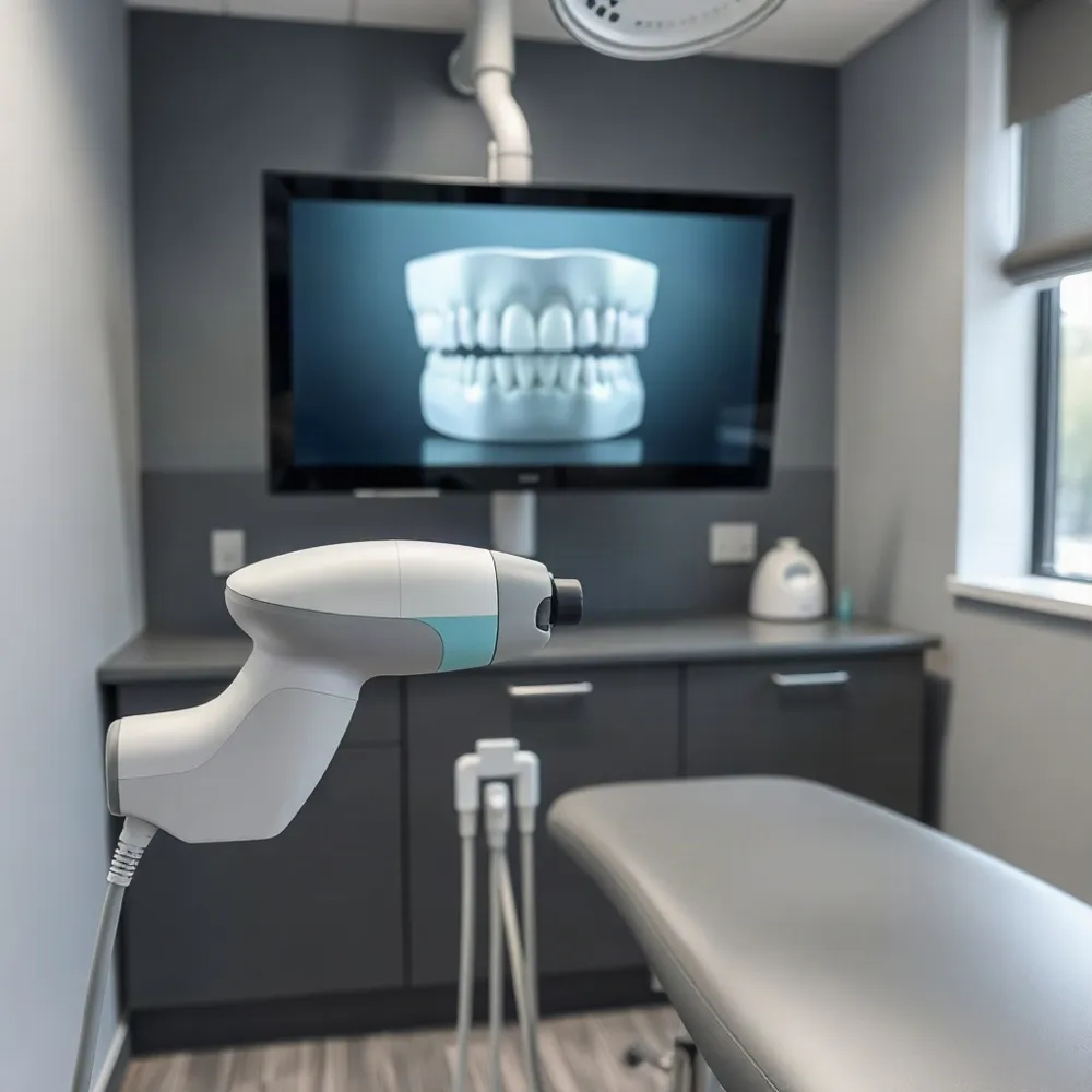 An intraoral scanner is a small handheld device that the clinician guides over the teeth and gums. It records views of the mouth’s surfaces without placing a tray of impression material around the teeth. This process creates a digital impression, a useful starting point for some treatment plans.
As the scanner moves, it directs light onto oral surfaces, and imaging sensors record the reflected information. Software aligns and combines the images to build a three-dimensional model. The method is described in this overview of intraoral scanners, though the clinical team’s judgment remains central to interpreting the result.
The developing model can appear on-screen during the appointment. The clinician can inspect the captured areas and, if a spot is missing or unclear, scan that area again before moving on. For patients, seeing the model may also make it easier to follow an explanation of the teeth or a proposed treatment.
A scan can often be completed in minutes, but the time varies with the area being recorded, access to the teeth, and conditions in the mouth. Saliva, movement, or limited access may affect image capture, so a scan is not necessarily equally simple in every case. For background on what patients can expect from a digital impression appointment, see what to expect from digital impressions.
At Ashley E. Burns, DDS in Midland, digital tools should be understood as part of an individualized dental evaluation, not as a substitute for a conversation about treatment needs. Whether a scan is suitable depends on the planned care and the clinician’s assessment.
Comfort Without Impression Material
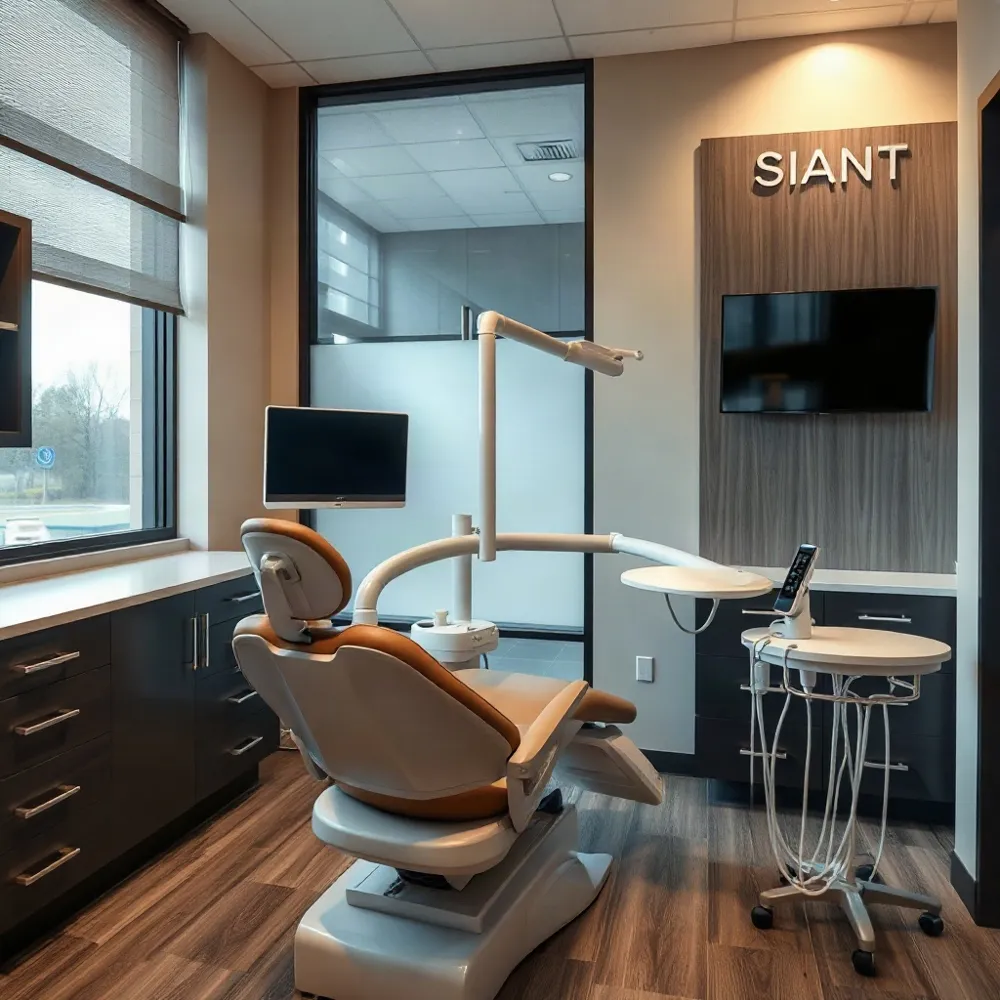 A conventional impression uses a tray filled with material that stays in the mouth while it sets. Some patients dislike the taste or the full feeling of the tray, and it can trigger gagging. An intraoral scan avoids that material, which may make the impression process more comfortable for people who find traditional molds unpleasant. Research summarized in a review found that patients generally rated scanning more favorably for several sensory experiences, including taste and nausea, though preferences vary.
A scan is not automatically comfortable for everyone. The wand may feel unfamiliar, its sound may be noticeable, and reaching the back teeth can feel awkward. Scanning can also take attention and cooperation, so the clinician may adjust the approach to suit the patient. Penn Dental Medicine’s overview of digital dentistry describes how scans can avoid the mess and gagging some people experience with conventional impression material.
Before starting, the dental team can explain what the scanner will do and describe the sequence, such as scanning one area at a time. Patients can ask questions or request a pause if they need a break. At Ashley E. Burns, DDS, patient-centered care means discussing the process and individual concerns rather than assuming one impression method will suit everyone. Patients who want to compare the approaches can read more about digital and traditional impressions.
Where Digital Impressions Can Help
(Where Digital Impressions Can Help):
A digital model can give the dental team a detailed record of the teeth and nearby tissues to use when planning care. For restorative work, that record may support the design of crowns and bridges, while orthodontic planning and some dental appliances can also benefit from a digital impression. The scan is one source of information, not a treatment decision on its own.
During a visit at ashleyburnsdds.com, a clinician can discuss the patient’s findings and proposed care as part of a personalized treatment conversation. When a digital model is available, viewing it together on-screen may make the shape of the teeth and the areas being discussed easier to understand. Penn Dental Medicine’s overview of digital dentistry describes this chairside use of scans to help explain treatment.
When a case involves a dental laboratory, a digital file can be sent electronically rather than mailing a physical impression. This may help the practice and laboratory coordinate, though it does not guarantee a shorter overall treatment timeline. A digital model can also support computer-aided design and manufacturing workflows for restorations, as described in research comparing digital and conventional impressions.
The right impression method depends on the planned treatment, the patient’s anatomy and needs, and the clinician’s assessment. Some cases may be well suited to scanning, while others may call for a conventional impression or a different approach. The dentist can explain why a method fits the case and how the resulting record will be used.
Reviewing the Scan Before You Leave
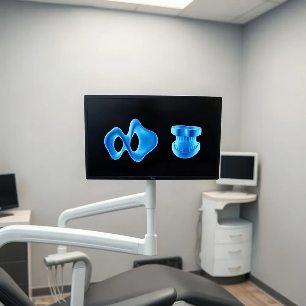 A digital impression can be checked while you are still in the chair. The clinician can inspect the model on screen for missing or unclear areas and, if needed, scan a localized spot again rather than starting over. This real-time review can make it easier to confirm that the information needed for planning has been captured.
A physical impression is checked after it has been taken. If it is distorted or incomplete, the dentist may need to take another full impression. Digital scanning does not remove every possibility of a retake, but reviewing the model during the appointment can help identify a problem before you leave. Research comparing intraoral scans with conventional impressions also notes that both methods have potential sources of error.
Once the scan is ready, its digital file can be sent to a dental laboratory without mailing a physical impression. That can support direct communication between the office and lab, but it does not guarantee a shorter wait for treatment. The lab’s schedule, the procedure, and any needed clinical steps still affect timing.
At Ashley E. Burns, DDS in Midland, reviewing a scan can be part of a clear conversation about what the images show and what happens next. A visual model may help you follow the discussion and ask questions, while the dentist’s examination and judgment guide the plan. As Penn Dental Medicine explains, showing a digital scan chair-side can support treatment discussions. Careful review may help the process feel more organized, though it cannot promise perfect results or fewer visits.
If you want to know whether a scan is suitable for your treatment, ask how the digital model will be reviewed and shared with the laboratory.
Accuracy Depends on the Case
How accurate are digital dental impressions made with intraoral scanners? They can provide accurate records for many restorations and may compare well with conventional impressions in appropriate cases. That does not mean a digital scan is always more accurate: the result depends on the scanner, the area being recorded, and the conditions in the mouth. Research comparing intraoral scans with conventional impressions also highlights that scanner technique can affect model accuracy.
An intraoral scanner builds a model by capturing images and joining them together in software. Each join can add a small deviation, so errors may accumulate across a longer scan. Clinicians also need a clear view of the relevant tooth surfaces. Saliva, limited access, reflections, or movement of teeth and soft tissues can make details harder to capture.
When an area is unclear or incomplete, the dentist may rescan it or change the approach. A conventional impression may be more suitable when the anatomy or treatment makes reliable scanning difficult. The appropriate method depends on the case, rather than on a general assumption that one technique is always preferable.
A scan is one source of information, not a substitute for clinical judgment. The dentist evaluates the captured details alongside the treatment needs and checks how the finished restoration fits. At Ashley E. Burns, DDS, patient-centered care and clear explanations are part of helping patients understand their options and what an impression can, and cannot, show.
When Another Approach May Fit Better
Digital scanning is highly effective in most cases, but certain factors like saliva, limited access to back teeth, or movement of the tongue and soft tissues can require adjustments. The clinician may need to dry an area, adjust the scan path, or scan part of the mouth again to capture the clearest details.
Some details, including deep edges below the gumline, can be difficult to record optically. A study comparing scanning with conventional impressions notes that digital models are assembled from overlapping images, and minor discrepancies can accumulate across a longer scan. Its findings highlight that accuracy depends on the case and technique, showing that each method has its strengths. Research on intraoral scanning and conventional impressions
Digital scanning requires suitable equipment, ongoing maintenance, and team training. Digital files may be sent electronically, but the handoff and review process depend on the practice’s workflow and its laboratory. The choice of impression method depends on the specific clinical situation and practice workflow.
Conventional impressions remain useful when a scan does not capture the required anatomy well or when the treatment calls for another approach. The choice should reflect the clinical details, the patient’s comfort, and the information needed for the restoration or appliance. The dentist can explain why one method is recommended and whether another option is appropriate.
At Ashley E. Burns, DDS, treatment decisions are grounded in patient-centered care. Because practice-specific scanning equipment and workflows vary, if a scan proves difficult, ask what information is missing, whether another scan or an impression is planned, and how the choice supports the intended treatment. What to expect from digital impressions
Choosing the Right Impression Method
Digital scans may feel more comfortable for patients who dislike impression material, and the on-screen model can help make treatment discussions easier to follow. Comfort and clinical suitability vary, so a scan is not automatically the best choice for every person or procedure. Penn Dental Medicine’s overview of digital dentistry describes how scans can support visual review and communication during care.
At Ashley E. Burns, DDS, treatment recommendations are grounded in comprehensive, patient-centered care. If impressions are part of your treatment, tell your dentist about gagging, anxiety, limited jaw opening, or other concerns. Ask which impression methods are available and how each fits your needs. A conversation about comfort and clinical goals can help you understand the options before deciding together.