
Seeing Your Smile More Clearly
An intraoral camera is a small handheld device that captures magnified images of teeth and gums and displays them on a screen. Looking at the same area together can make a dentist’s explanation more concrete, especially when a finding is hard to see in a mirror. Research describes these images as a useful aid for patient education and shared treatment discussions, sometimes called co-diagnosis (clinical review of intraoral cameras).
At Ashley E. Burns, DDS in Midland, visual technology supports patient understanding, including the use of intraoral cameras to help patients see what the dental team sees (advanced dental technology and treatment comfort). Images provide only a view of visible surfaces, however. They do not replace the dentist’s full examination or other tests that may be needed to assess a concern.
How Intraoral Cameras Show Findings
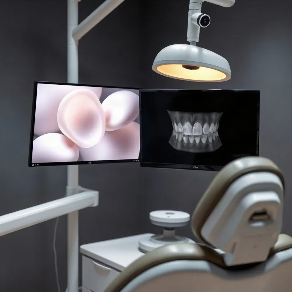 An intraoral camera is a small handheld device with a camera and light. It captures magnified images or video of visible teeth and oral tissues, then displays them on a screen. Unlike a mirror view, the image can be enlarged so the dentist can point out a specific area while you see the same view.
That shared view can make a conversation about oral health more concrete. A dentist might use an image to discuss plaque, inflamed gums, tooth wear, a fracture, or a possible defect in a filling or crown. What appears depends on the image quality, camera, and area being viewed, so a mark on screen does not by itself establish a diagnosis or determine treatment.
Reviewing personal images can support patient education and shared treatment discussions, sometimes described as co-diagnosis. A research review describes these uses, while also noting that evidence varies across applications and that images can produce false-positive findings in some settings. The image is one aid to the dentist’s assessment, not a substitute for clinical judgment. The review of intraoral camera applications discusses both the educational value and limitations.
A camera shows visible surfaces; it does not reveal the inside of a tooth or the supporting jaw structures that may be assessed with X-rays. The dentist considers the image alongside an examination, symptoms, and any other tests needed. If a tooth hurts despite looking unremarkable on screen, be sure to describe the pain and when it occurs.
Images may be saved in the dental record, reviewed at a later visit, or compared over time to document visible changes. With appropriate consent and privacy safeguards, they may also support a consultation with another dental professional. Educational information on ashleyburnsdds.com describes how seeing what the dentist sees can help reduce uncertainty; ask your dental team whether images are being saved and how they are used.
What Images Can and Cannot Tell You
 A close-up image can make a dentist’s observations easier to follow. Looking together at areas where plaque collects or gums appear inflamed can also help clarify brushing and flossing guidance. Research describes intraoral cameras as tools for patient education and shared discussion, not stand-alone diagnostic tests (clinical review of intraoral cameras).
When images are saved, comparing them at later visits may help the dentist and patient review visible changes in teeth, gums, or restorations. The images add context to an examination, but a photograph by itself cannot confirm a diagnosis or determine which treatment is appropriate. At Ashley E. Burns, DDS, image-based explanations fit within a broader conversation about observations and care options, rather than replacing the dentist’s assessment.
A camera records only the surfaces it can reach and show clearly. Areas between teeth may be difficult to view, and a surface image cannot reveal the inside of a tooth or supporting bone. A clear-looking picture therefore does not rule out every concern, and the dentist may recommend other tests when the examination or symptoms call for them.
Patients can ask what a particular image shows, whether the finding needs monitoring or further testing, and what information supports a recommendation. If images are captured, stored, or shared, patients should also be able to ask how consent and confidentiality are handled. The clinical review stresses appropriate consent and privacy safeguards for patient images.
How Intraoral Scanning Builds a Model
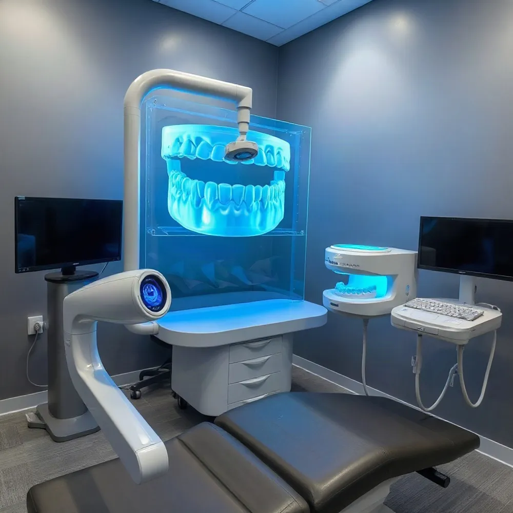 An intraoral camera and an intraoral scanner may both look like handheld wands, but they capture different information. A camera takes photos or video of visible areas, while a scanner records the shape of teeth and gums to form a digital impression. For a closer look at how scanning differs from photography, see how intraoral scanning builds a model.
To create a model, the clinician moves the scanner over the teeth and nearby tissues. The device projects light and records the reflections from many overlapping views. Software aligns and processes those views into a developing three-dimensional model on screen. The clinician can check which areas have been captured and take more views if data appears missing or unclear.
A digital impression can support treatment planning and, when appropriate, be shared with a dental laboratory to help design restorations or appliances such as crowns, bridges, or aligners. Scanning also allows on-screen review and correction during capture, and digital files avoid some potential distortion associated with impression materials and their handling.
Some patients may find scanning less messy than a tray impression, but comfort varies. The best method depends on the patient, the area being recorded, and the treatment. Ashley E. Burns, DDS, emphasizes patient education through digital imaging, where images can be reviewed and discussed with the dentist. That does not establish that the practice offers intraoral scanning, so patients can ask which impression options are available for their care.
When a Scan Needs Another Approach
An intraoral scanner can build a useful digital model, but it records only the surfaces it can see clearly. Tight spaces between teeth, the back of a tooth, or a margin close to the gumline may be difficult to capture when access is limited.
Saliva, blood, reflections, and moving soft tissue can obscure details. Scanning technique also matters: the team may need to adjust the wand’s path, keep the area dry, or gently move tissue aside. If parts of the model look unclear or incomplete, they may take more images or repeat the scan.
For care at ashleyburnsdds.com, the appropriate imaging approach depends on the area being treated and the conditions in the mouth, not on choosing digital impressions in every case. A conventional impression remains an option when it is better suited to capturing the information needed. The dentist can explain the reason for the method selected, including how it supports the planned care. For more on scan access challenges, see this step-by-step intraoral scanning technique.
A Shared View of Care
Looking at an intraoral image together can make a conversation about visible changes clearer and more collaborative. Research describes this shared review as a way to support patient education and treatment discussions, while emphasizing that images are an aid to the dentist’s assessment, not a diagnosis on their own (review of intraoral camera applications).
At Ashley E. Burns, DDS in Midland, visual explanations fit a patient-centered approach to care. Ask what an image shows, whether further examination is needed, and what options are appropriate. If a digital impression or another imaging method is discussed, ask how it differs from a camera image and what information it can provide.