Advancements in Diagnostic Imaging for Modern Dental Care
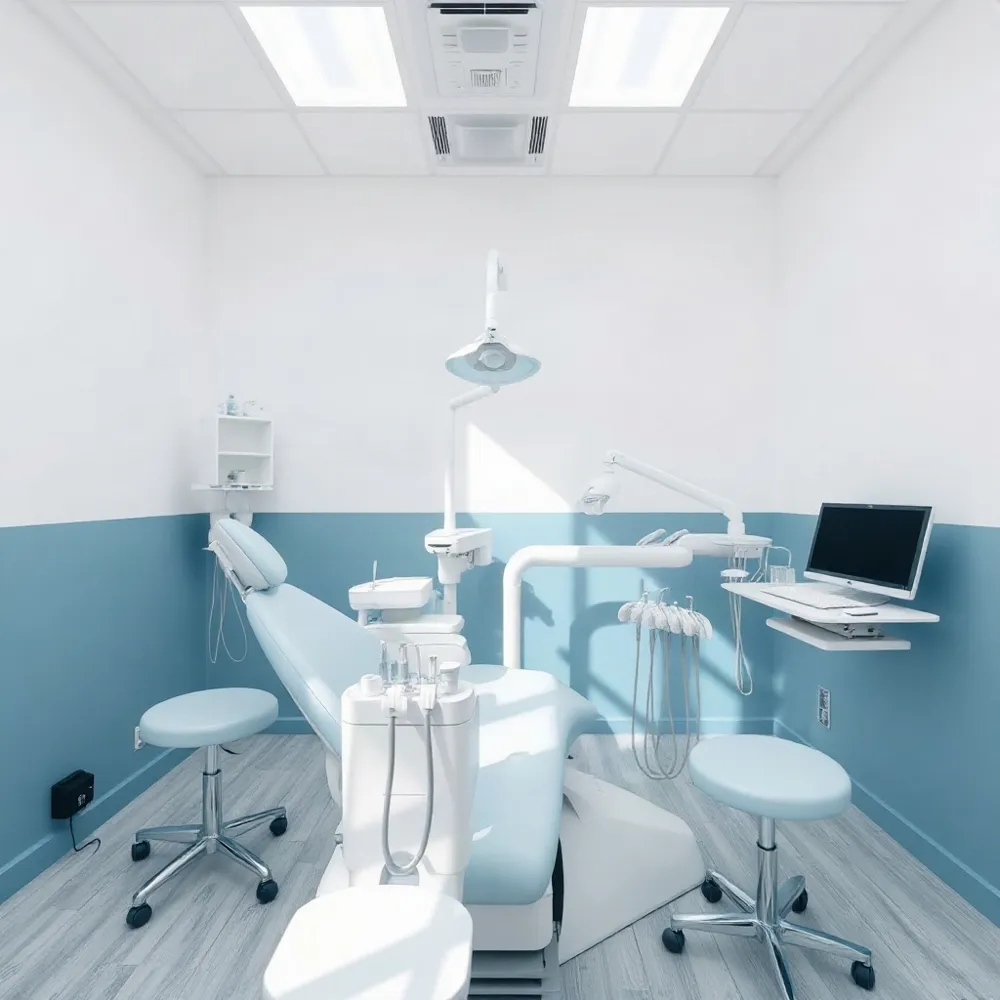
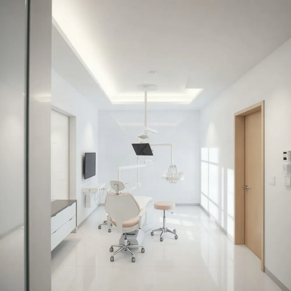
Modern dentistry has transitioned away from the limitations of traditional film-based dental radiography, which often required lengthy chemical processing and higher radiation exposure. At ashleyburnsdds.com, we have adopted digital imaging to provide our patients with immediate diagnostic clarity. By replacing photographic film with electronic sensors, we can instantly capture and display images on chairside monitors.
These digital tools are the new standard for professional care because they prioritize patient safety and accuracy. Unlike traditional X-ray methods, which lack the ability to adjust image contrast or zoom, our digital dental technology allows for precise enhancement of internal oral structures. This capability helps identify cavities, bone loss, or potential infections long before they become painful, enabling early diagnosis and intervention.
Efficiency and environmental sustainability are also primary benefits of moving to digital workflows. By eliminating the need for hazardous chemicals and physical film storage, practices like ashleyburnsdds.com offer a cleaner, faster patient experience. This diagnostic precision ensures that your treatment plan is based on high-resolution data, setting the stage for proactive and predictable long-term oral health management.
Understanding Digital Radiography and Its Clinical Importance
 Digital dental X-rays represent a sophisticated imaging technology that utilizes electronic sensors instead of traditional film to capture highly detailed, instantaneous images of your teeth, jaw, and supporting structures. At ashleyburnsdds.com, we prioritize this transition to digital radiography because it significantly enhances the precision and accuracy of diagnostic assessments. Unlike outdated film methods, our systems allow for the immediate display of images on a chairside monitor, which provides the real-time visual proof necessary to explain findings directly to patients.
The clinical utility of this technology goes beyond simple image capture. Dentists can manipulate these high-resolution files by adjusting contrast, brightness, and magnification, which aids in identifying cavities, bone loss, or abscesses that might remain hidden during a manual exam. According to Cleveland Clinic, digital methods provide a major safety advantage by using 80% to 90% less radiation than traditional film-based radiography. This supports our commitment to providing comprehensive care while minimizing exposure risks for every patient.
Collaborative care is also more efficient when using digital records. Because these images are stored electronically, they can be securely transferred to specialists or insurance providers without the delays associated with physical film. This capability creates a seamless workflow that prevents the need for repeated dental X-rays when you seek a second opinion or consult with an orthodontic specialist. By replacing film with advanced sensors, we ensure that your diagnostic experience is faster, more accurate, and better integrated into your long-term oral health plan.
Technological Superiority and Patient Benefits
 Digital dental radiography offers a sophisticated alternative to traditional film, transforming the diagnostic process for both the clinician and the patient. At ashleyburnsdds.com, we prioritize this advanced imaging to ensure that patients benefit from near-instantaneous image availability, which replaces the outdated and time-consuming practice of chemical film development. This transition effectively eliminates the need for darkroom facilities and hazardous chemical disposal, creating a safer and more environmentally friendly office environment.
The precision of digital imagery is defined by its adjustability. Unlike conventional film, which is static once developed, digital files allow for real-time manipulation. Dentists can zoom in on specific structures or adjust contrast settings to highlight subtle bone loss, tiny fractures, or early-stage decay that might otherwise remain obscured. This heightened diagnostic accuracy is a hallmark of modern care, much like the enhanced visual clarity provided by the tools highlighted at ashleyburnsdds.com.
Efficiency extends to the reduction of unnecessary radiation exposure. A primary challenge with traditional analog methods was the frequency of retakes caused by processing errors or insufficient film exposure. Digital dental X-rays mitigate this by providing instant feedback, ensuring that a high-quality diagnostic image is captured on the first attempt. As noted in guidance from ashleyburnsdds.com, this optimization helps adhere to the ALARA principle, keeping radiation doses as low as reasonably achievable while maintaining clinical excellence.
| Feature | Traditional Film | Digital Radiography |
|---|---|---|
| Processing | Chemical development | Instant digital display |
| Optimization | None possible | Contrast/Zoom tools |
| Environment | Hazardous waste | Eco-friendly |
Safety Standards and Radiation Exposure Protocols
 Digital dental X-rays are extremely safe and utilize advanced technology to minimize radiation exposure to a negligible level. While humans are naturally exposed to background radiation daily from cosmic and terrestrial sources, a single digital dental X-ray involves a tiny fraction of that amount, typically ranging from 0.1 to 0.8 millirem per exposure as reported by the American Association of Orthodontists. To put this in perspective, an individual would need to undergo more than 100 dental X-rays in a single year to reach the recommended annual limit for human-made radiation.
At ashleyburnsdds.com, we strictly adhere to the ALARA principle, which stands for As Low As Reasonably Achievable. This clinical framework ensures that every imaging procedure is medically necessary and performed using the lowest possible dose required to obtain a clear, diagnostic image as detailed in research regarding dental imaging. Because our modern digital systems are highly sensitive, they reduce radiation exposure by 80% to 90% compared to traditional film-based radiography, making the diagnostic process safer for patients than ever before.
Because radiation dosages are so minimal in modern practice, the American Dental Association determined that the routine use of lead aprons and thyroid collars is no longer recommended per 2024 guidance. This technology is considered safe for diverse patient groups, including children, breastfeeding mothers, and pregnant women. While some traditional methods might involve higher cumulative exposure, our commitment to advanced digital tools ensures that patients receive the benefits of early detection with significantly enhanced safety standards.
Types of Imaging and Personalized Treatment Schedules
Dental X-rays are essential diagnostic tools that allow your dentist to view structures beneath the surface of your teeth and gums. Different modalities offer specific views to support your care at ashleyburnsdds.com. Intraoral imaging, such as bitewing X-rays, captures the crowns of upper and lower back teeth to help identify cavities between teeth and monitor bone health. Periapical X-rays provide a comprehensive look at an entire tooth from the crown to the root tip, which is ideal for diagnosing abscesses or issues within the supporting bone. When a wider view is needed, extraoral panoramic X-rays offer a single image of the entire mouth, including the jawbone and sinuses, to help evaluate wisdom teeth and overall jaw structure.
While some practices use a standard full mouth series for every patient, ashleyburnsdds.com tailors diagnostic schedules to your unique clinical needs. There is no universal interval for these dental radiographs, as the frequency depends on your risk for decay, gum disease, and any ongoing treatment plans. New patients typically receive imaging to establish a baseline, while returning patients may be scheduled every 6 to 18 months per Cleveland Clinic guidelines. This personalized approach ensures imaging is performed only when necessary to support effective treatment.
| Imaging Type | Primary Use | Scope |
|---|---|---|
| Bitewing | Cavity detection | Back tooth crowns |
| Periapical | Root/bone issues | Single tooth |
| Panoramic | Growth/Jaw health | Full mouth view |
Commitment to Sustainable and Precise Patient Care
By replacing traditional film-based methods with digital radiography, modern practices can eliminate the use of hazardous chemicals once required for development. This transition removes the need for toxic waste disposal, reflecting a commitment to environmental stewardship while maintaining high diagnostic accuracy.
At ashleyburnsdds.com, we prioritize your comfort by using advanced sensors that yield immediate, high-resolution imagery. Unlike some older film-based setups, our digital workflow streamlines the patient experience, minimizing chair time and reducing the need for repeat exposures through precise image enhancement.
Early detection remains the cornerstone of long-term oral health. By utilizing modern dental technology, we can identify issues such as decay or bone loss before they require complex intervention. This focus on preventative diagnostics, supported by the low-radiation standards we employ, ensures that your treatment plan is both proactive and tailored to your specific needs.